Insulin-derived growth factors mainly include insulin-like growth factor 1 (IGF1) and insulin-like growth factor 2 (IGF2), which are core ligands of the IGF axis, regulating embryonic development, organismal growth, cell proliferation and survival, and metabolic homeostasis. These two IGFs exert opposite growth-regulating effects through different IGFRs: IGF1 promotes growth through IGF1R, while IGF2 inhibits growth through IGF2R. Abnormal activation of this pathway drives tumorigenesis, metastasis, and drug resistance, making it a highly promising target for tumors, metabolic diseases, and developmental disorders.
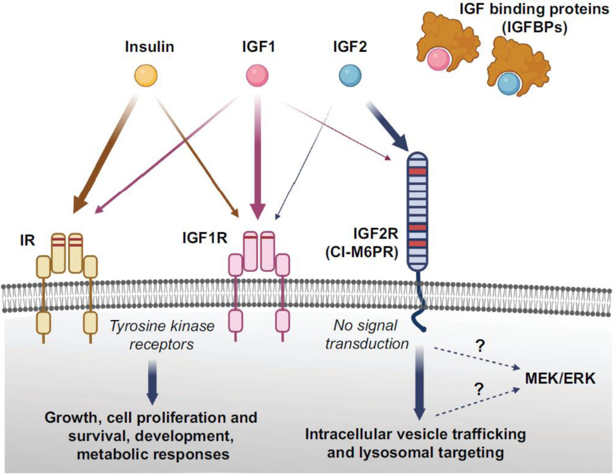
(Data source: Alberini CM. Trends Neurosci. 2023)
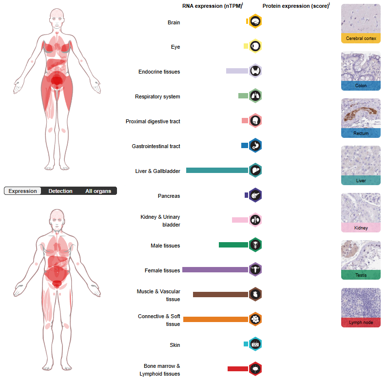
IGF1 and IGF2 expression distribution
IGF1 is mainly synthesized by the liver and secreted into the blood as a circulating endocrine factor, regulated by growth hormone (GH). It is also expressed in various tissues such as bones, cartilage, muscles, kidneys, lungs, and brain tissue. It is continuously expressed after birth and is a major regulator of the body's growth after birth.
IGF2 is primarily expressed at high levels in embryonic/fetal tissues, and can be detected in the liver, lungs, skeletal muscle, and placenta during embryonic development. In adulthood, its expression is downregulated in most tissues, but it can be reactivated in some epithelial, mesenchymal, and tumor tissues.
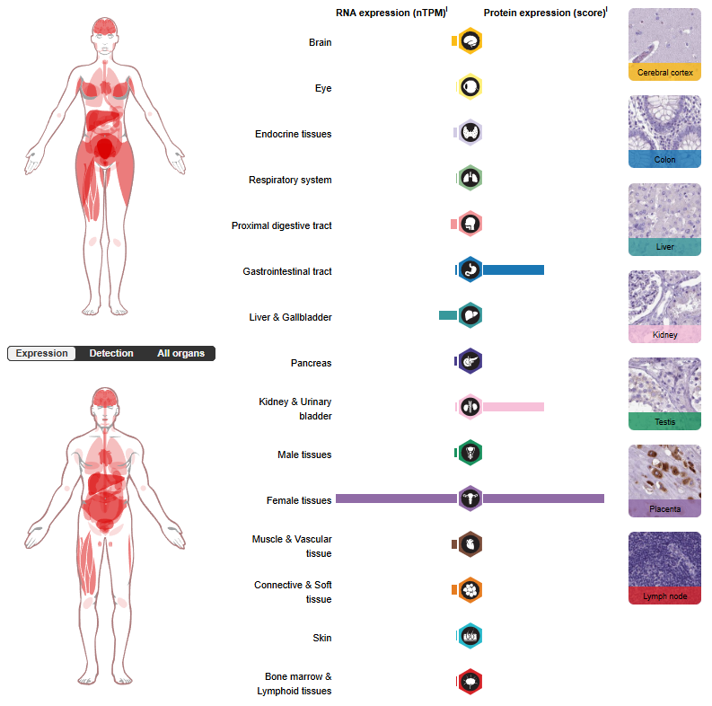
(Data source: uniprot)
The structure and receptors of IGF1 and IGF2
IGF1 and IGF2 are single-chain polypeptides that are highly homologous to insulin precursors. The mature protein contains four domains: B, A, C, and D.
IGF1: Mature IGF1 consists of 70 amino acids with a molecular weight of approximately 7.6 kDa. Its B and A domains are conserved and homologous to the insulin A/B chain. The C domain is a variable linker peptide connecting the B and A domains, equivalent to the C peptide of proinsulin, but IGF-1 can be secreted without cleaving the C domain. The D domain, located at the C-terminus, is absent in insulin and participates in receptor binding and IGFBP interaction.
IGF2: Mature IGF2 consists of 67 amino acids with a molecular weight of approximately 7.5 kDa. Its sequence is about 70% homologous to IGF1, and it also possesses B/A/C/D domains. The A domain is homologous to the insulin A chain and contains a key α-helix; residues in the A domain (such as A54 and L55) are involved in IGF2R binding specificity. The C region is shorter than that of IGF1, which is the structural basis for the difference in receptor affinity. The D domain is located at the C-terminus and is homologous to �the D domain of IGF1, but its sequence is different.
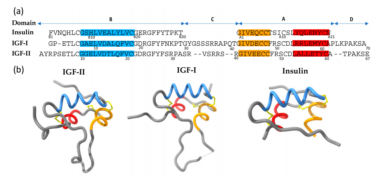
(Data source: Blyth AJ, et al. Cells. 2020)
IIGF-related receptors include the insulin receptor (IR), which exists in two molecular forms, isoform A (IR-A) and isoform B (IR-B); the IGF1 receptor (IGF1R) and the IGF2 receptor (IGF2R, also known as the mannose-6-phosphate receptor, IGFR2/M6FR).
IGF1R is the main receptor for IGF1 and IGF2. IGF1 has the highest affinity for IGF1R, followed by IGF2.
IGF2R (M6PR) is a single-stranded transmembrane protein with no intracellular kinase activity. It preferentially binds to IGF2, mainly mediates the internalization and degradation of IGF2, and negatively regulates IGF2 levels.
Insulin receptor A subtype (IR-A): IGF2 can bind to IR-A with high affinity, mediating proliferation and anti-apoptotic signals in tumor cells, which is a bypass mechanism for IGF2 to promote cancer.
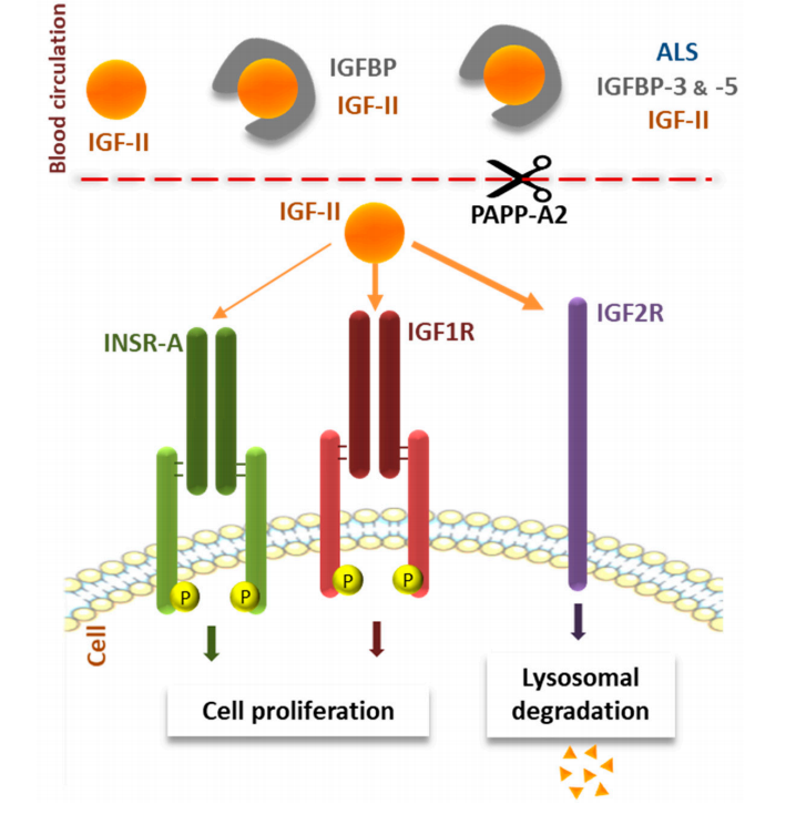
(Data source: Sélénou C, et al. Cells. 2022)
IGF1 and IGF2 signaling pathways, regulation, and their roles in tumors
Upon binding of IGF1/IGF2 to IGF1R (or IR-A), a conformational change in the receptor is induced, leading to tyrosine autophosphorylation of the intracellular β subunit. This recruits and phosphorylates the IRS (insulin receptor substrates, IRS1/IRS2), initiating two core downstream pathways:
The PI3K/AKT/mTOR pathway: Phosphorylation of IRS activates PI3K, catalyzing the conversion of PIP2 to PIP3, and recruiting PDK1 to activate AKT; AKT further inhibits the TSC complex and activates mTOR. This pathway inhibits apoptosis, promotes cell survival, protein synthesis, metabolic reprogramming, and enhances tumor cell drug resistance.
RAS/MAPK (ERK) pathway: The IRS/Grb2-SOS complex activates RAS, which in turn activates Raf , MEK , and ERK. This promotes transcription, cell cycle progression, proliferation, epithelial-mesenchymal transition (EMT), and invasion and metastasis (PMC).
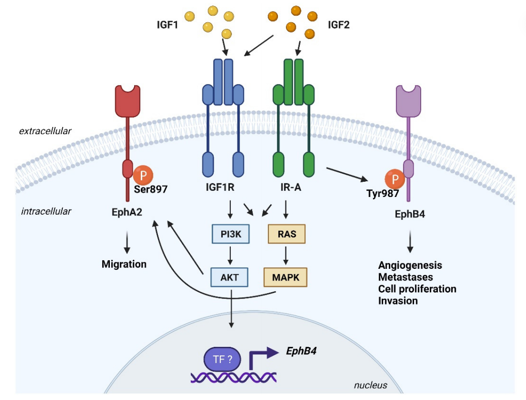
(Data source: Mancarella C, et al. Cells. 2021)
When IGF2 binds to IGF2R, it initiates the ligand clearance pathway, transporting IGF2 to lysosomes for degradation and inhibiting signaling; IGFBP can capture free IGF, block ligand-receptor interactions, and dynamically regulate pathway strength.
IGF2 exerts its effects in the tumor microenvironment by suppressing anti-tumor immunity. IGF2 inhibits T-cell immunity and promotes tumor inflammation by promoting the maturation of dendritic cells (DCs) and inhibiting their apoptosis, thereby causing them to secrete various cytokines. Furthermore, immunosuppressive cells increase the expression of immune checkpoint proteins such as PD-1/PD-L1 and CTLA-4, inhibiting T-cell activation and blocking antigen presentation. The expression of arginase 1 (ARG1), indoleamine 2,3-dioxygenase (IDO), and differentiation inhibitory factor 1 (Id1) increases the production of reactive oxygen species (ROS) and IGF2, and induces an immunosuppressive phenotype. T-cell exhaustion and the induction of an immunosuppressive phenotype can synergistically promote tumor growth.
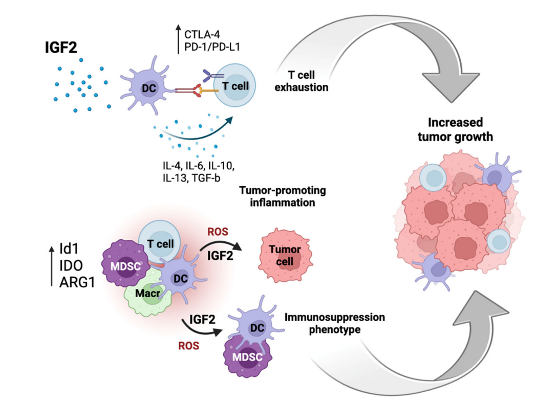
(Data source: Belfiore A, et al. Biomedicines. 2023)
Targeted therapy of IGF1 and IGF2
Xentuzumab is a humanized monoclonal antibody. It targets IGF-1/2 and blocks IGF proliferation/anti-apoptotic signaling. Xentuzumab inhibits the ability of both IGF ligands to interact with IGF-1R and may block IR-A compensatory signaling associated with IGF-1R antibody resistance by neutralizing IGF-2, which has a high affinity for IR-A. In cell-based assays, xentuzumab showed potent inhibition of both IGF-1R and IR-A signaling. Conversely, IGF-1R antibodies had no effect on IR-A signaling via IGF-2 and were less effective than xentuzumab in inhibiting IGF-1R signaling. In a bone metastatic prostate cancer model, IGF neutralizing antibodies effectively inhibited receptor activation of IGF-1 and 2 and reduced tumor growth. Furthermore, unlike IGF-1R antibodies, xentuzumab is less likely to disrupt growth hormone homeostasis and rarely induces hyperglycemia. In two phase I studies in patients with advanced solid tumors, xentuzumab monotherapy has shown preliminary antitumor activity. A phase Ib/II study in patients with HR-positive, HER2-negative breast cancer demonstrated that xentuzumab in combination with everolimus and exemestane was well-tolerated. A phase Ib trial evaluated the efficacy of the insulin-like growth factor 1/2 neutralizing antibody xentuzumab in combination with abemaciclib and endocrine therapy (ET) in patients with advanced/metastatic solid tumors, including hormone receptor (HR)-positive breast cancer. The combination therapy of xentuzumab, abemaciclib, and ETs demonstrated manageable tolerability and encouraging efficacy, particularly in patients with breast cancer and non-visceral metastases.
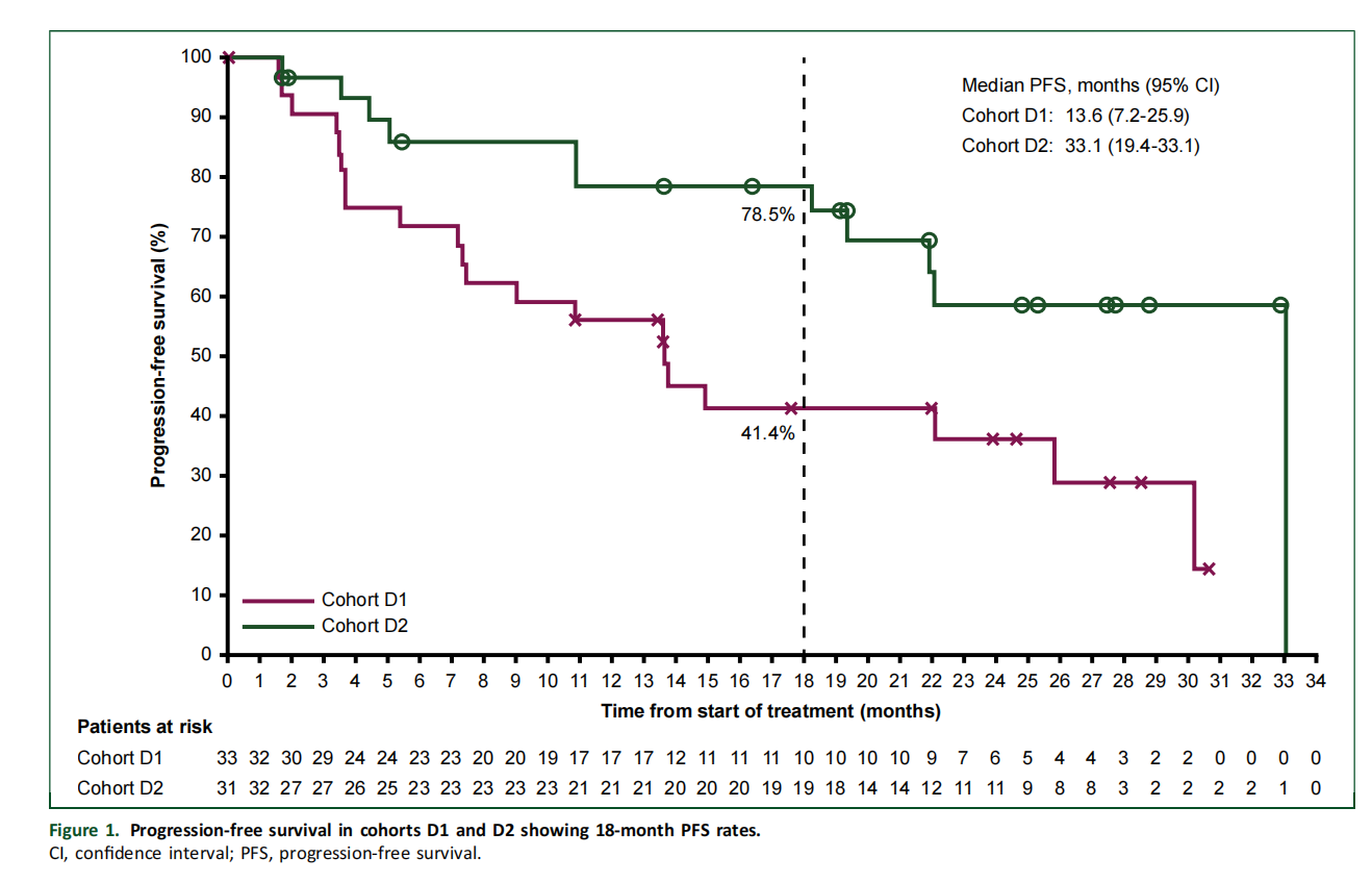
(Data source: Yee D, et al. ESMO Open)