BK polyomavirus (BKPyV) is a member of the Polyomaviridae family (double-stranded DNA viruses), a small, non-enveloped DNA virus that is ubiquitous in the population and remains latent in the body. It can reactivate in individuals with compromised immune systems, particularly after kidney transplantation, and can activate various transcription factors and immune mediators. Although reactivation is usually asymptomatic, it can manifest as acute kidney injury (AKI), a risk factor for early transplant organ loss. Immune responses to BKPyV are crucial in controlling the virus and protecting organs from damage during infection. The presence of BKPyV in kidney transplant recipients (KTRs) most commonly induces urinary tract infections (UTIs), which are the most common cause of AKI in KTRs. Currently, there are no effective antiviral drugs for BKV infection, and the primary approach to managing viral reactivation is reducing immunosuppression. Developing immunotherapy-based therapies to combat BKV plays a vital role.
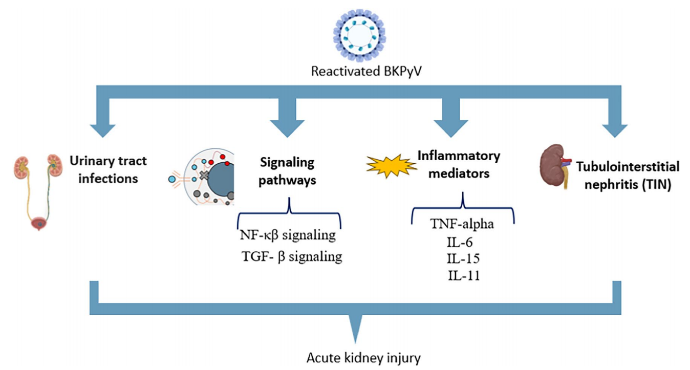
(Data source: Bizhani S, et al. Virol J. 2025)
The structure of BKV and VP1 proteins and their receptors
Members of the Polyomaviridae family share structural similarities, exhibiting similar capsid size, high genetic homology, and comparable genome size. BKV possesses a small, non-enveloped icosahedral capsid, 40 to 44 nanometers in diameter, encoded by the viral capsid proteins VP1, VP2, and VP3. These capsid proteins surround a single DNA molecule that binds to histones to form a chromatin chain. The capsid proteins are arranged in a T=7 d icosahedral structure, containing 360 VP1 molecules organized into 72 pentamers. Each pentamer contains individual portions of the two minor capsid proteins, VP2 and VP3. Therefore, VP1 is the only viral protein exposed on the viral particle surface, responsible for binding to host cell receptors, thereby facilitating viral entry into the cell.

(Data source: Hurdiss DL, et al. Structure. 2018)
VP1 is a protein composed of 362 amino acids (42 kDa) divided into five rings, designated BC, DE, EF, GH, and HI, which connect different parts of the polypeptide chain. As previously mentioned, VP1 forms pentamers, which are present on the outer surface of the viral capsid. The importance of the VP1 rings in mediating capsid assembly has been demonstrated. The pentamer consists of a ring structure composed of five β-barreled VP1 monomers, tightly linked by loops that interact between the β-chain frameworks. The N-terminal region of VP1 is located inside the viral particle and mediates DNA binding. The C-terminal subunit of each VP1 monomer forms an arm extending to the adjacent cap body monomer, binding them together to form the viral particle.
Each VP1 pentamer interacts with either an internal protein, VP2 (351 amino acids; 38 kDa) or VP3 (232 amino acids; 27 kDa), which inserts into the central cavity in a hairpin-like manner via hydrophobic interactions. Both proteins are expressed by the same late mRNA transcript and share a C-terminal amino acid sequence, but VP2 has a unique N-terminal amino acid sequence, including a presumed myristoylation site at Gly-2. However, modification of this site by BKPyV VP2 has not been detected by mass spectrometry. The shared C-terminal fragment of VP2 and VP3 contains important features, including the VP1 binding region, DNA binding region, and NLS. Neither protein is essential for viral assembly, and their removal does not affect the stability of viral particles.

(Data source: Hurdiss DL, et al. Structure. 2018)

(Data source: Pietrobon S, et al. Front Immunol. 2017)
Mechanism of BKV entry into cells
Unlike other polyviruses, BKV enters host cells via caveolae-mediated entry, while other polyviruses typically rely on vesicle protein-mediated endocytosis. The BKV VP1 protein has a cleft between the β-chain C1 (BC1) and BC2 loops, allowing it to bind to the α2-8 linked sialic acid motif in the galactosides GD1b and GT1b expressed on the host cell membrane. Subsequently, the virus enters the cell via caveolae-mediated endocytosis. Once inside the cell, BKV is transported to the endoplasmic reticulum via microtubules and undergoes capsid uncoating via the classical endocytic pathway. Following VP1 uncoating, VP2 and VP3 mediate BKV entry into the nucleus via introduction proteins. After entering the nucleus, the BKV genome remains free in human cells; in contrast, in rodent cells, the BKV genome integrates into the host DNA, leading to rodent tumors.

(Data source: Ambalathingal GR, et al. Clin Microbiol Rev. 2017)
BKV targeted therapy
Potravitug is a monoclonal antibody targeting the VP1 outer membrane protein of the BK virus. Its mechanism of action involves blocking viral binding and entry into host cells, thereby preventing infection and inhibiting viral replication. Developed by Memo Therapeutics AG, later acquired by Ipsen, Potravitug received Fast Track designation from the U.S. Food and Drug Administration (FDA) in May 2023 and Orphan Drug Designation in the European Union in December 2025. The Phase II SAFE Kidney II trial is currently the largest placebo-controlled clinical trial for the treatment of BKPyVAN in kidney transplant patients, enrolling 95 patients from 22 research centers in the United States. The primary results showed that Potravitug was significantly effective: at week 20, the proportion of patients with ≥1-log10 viral load reduction or undetectable viral load was higher than in the placebo group, and the histological characteristics of BKPyVAN were also improved. Overall data indicate that Potravitug has significant clinical value, providing sustained and significant antiviral effects and reducing the incidence of BKPyVAN.
Traxivitug (MAU868) is a human IgG1/λ antibody isolated from memory B cells of a healthy, BKPyV seropositive donor initially identified during screening for cells capable of binding BKPyV VLPs (types I and IV). The MAU868 scFv binds to an epitope located on the outer surface of the BKPyV VP1 pentamer. The interacting surface consists of several continuous and discontinuous (i.e., non-continuous) sequences: residues 77–80, 169–186, and 191–192. These residues constitute a 3-dimensional conformational epitope recognized by MAU868, with tyrosine 169 (Y169), arginine 170 (R170), and lysine 172 (K172) providing crucial contact interactions. The FIH clinical trial results demonstrated that a single dose of MAU868 (up to 100 mg/kg intravenously) was safe and well-tolerated, and provided human data to guide dosage and regimen selection in subsequent studies. MAU868 holds promise as a first-in-class antiviral drug for the treatment and/or prevention of BKPyV disease.

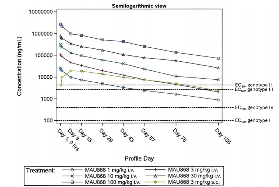
(Data source: Abend JR, et al. Am J Transplant. 2024)