COVID-19, caused by SARS-CoV-2, has evolved from a devastating global pandemic over the past five years into an endemic disease. Despite the widespread availability of vaccines, COVID-19 continues to pose a threat to public health, with the emergence of novel viral variants. SARS-CoV-2 relies on its essential receptor, angiotensin-converting enzyme 2 (ACE2), to enter cells. The trimeric spike protein (S protein) encoded by the virus mediates SARS-CoV-2 entry into host cells and induces a strong immune response, making it an important target for the development of therapeutics and vaccines.

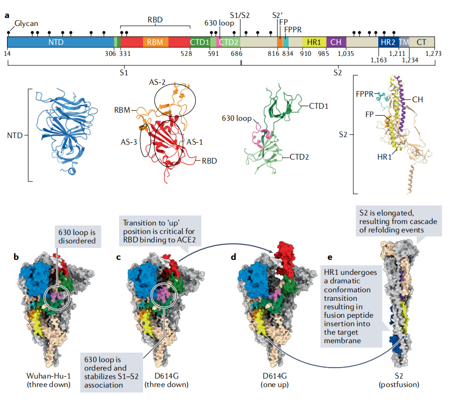
(Data source: Jackson CB, et al. Nat Rev Mol Cell Biol. 2022)
The structure of the SARS-CoV-2 spike protein and its receptor
The protein sequence of the original Wuhan strain of SARS-CoV-2, Hu-1, contains 1273 amino acid residues and 22 N-linked glycosylation sites. The monomeric S protein of SARS-CoV-2 is a type I membrane protein; each S protein trimer contains 66 N-linked glycans and belongs to the so-called type I viral fusion proteins, represented by influenza virus hemagglutinin proteins. This S protein is anchored to the viral membrane through its transmembrane region, presenting a large outer domain on the surface of the viral particle. S1 includes an N-terminal domain (NTD), a receptor-binding domain (RBD), and two C-terminal domains (CTD1 and CTD2), while S2 includes FP, heptameric repeat 1 (HR1), a central helix (CH), a linker domain (CD), heptameric repeat 2 (HR2), a transmembrane segment (TM), and a cytoplasmic tail (CT).
N-terminal domain (NTD): The NTD mainly consists of four stacked β-sheets and several connected flexible rings with several N-linked sugar groups.
The receptor-binding domain (RBD): consists of a core structure containing a five-stranded antiparallel β-sheet flanked by short connecting α-helices and an extended loop (the receptor-binding motif (RBM)) that binds tightly to one side of the core structure, forming the receptor-binding surface. In its downconformation, the RBD covers the central helical bundle of S2 and two other RBDs within the S protein trimer, while also resting against CTD1 on the same polypeptide chain and the NTD of an adjacent subunit. This conformation partially obscures the RBM, preventing it from binding to ACE2. When the RBD flips up, the adjacent CTD1 and NTDs move aside to accommodate the RBD's movement, thus fully exposing the RBM. These two CTDs are primarily composed of β-structures. There are three main non-overlapping antigenic sites on the RBD.
C-terminal domain (CTD): The CTD is primarily composed of β-structures. If the RBD is considered as an insertion between two antiparallel β-chains in CTD1, then CTD1 can also be considered as an insertion between two β-chains in CTD2. When the RBD adopts an " up " conformation, CTD1 is located below the RBD and rotated outward. CTD2 contains two stacked four-chain β-sheets. One of the β-sheets has an interchain loop containing an S1/S2 cleavage site, and one of the chains is the N-terminal fragment of S2. Therefore, the dissociation of S1 from S2 requires disrupting the stability of CTD2, thereby releasing the N-terminus of S2.
ACE2 is an 805-amino acid carboxypeptidase that removes a single amino acid from the C-terminus of its substrate. Previously identified as a receptor for other coronaviruses, it is the primary entry receptor for SARS-CoV-2, facilitating viral entry by binding to the RBD. It is a type I membrane glycoprotein containing an extracellular domain with metallopeptidase activity, serving as a key negative regulator of the renin-angiotensin system (RAS) for modulating vascular function. The peptidase active site does not overlap with the RBD binding site, and this enzyme activity is not essential for its receptor function. The interface between the S protein RBD and ACE2 is primarily composed of a slightly concave outer surface extending the RBM and the N-terminal helix of the receptor. ACE2 has 20 residues, and the RBD has 17 residues, forming a hydrophilic side-chain interaction network.
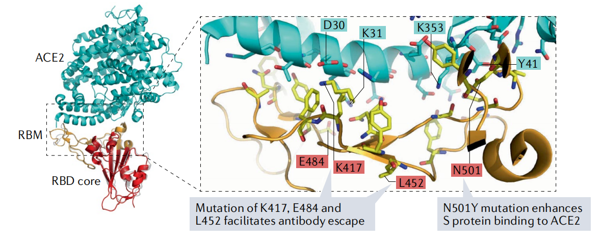
(Data source: Jackson CB, et al. Nat Rev Mol Cell Biol. 2022)
Mechanism of SARS-CoV-2 entering cells
The primary pathway of SARS-CoV-2 infection begins with the binding of the S protein to the ACE2 receptor on the surface of target cells. This interaction induces extensive conformational changes in the S1 and S2 subunits, bringing the viral envelope into close contact with the host cell membrane and exposing the internal S2′ cleavage site. Cleavage at this site is primarily mediated by transmembrane serine protease 2 (TMPRSS2) on the cell membrane or by soluble cathepsin L in the endosomal lumen following clathrin-mediated endocytosis (CME). This cleavage event leads to the externalization of the viral protein (FP) and its integration into the host cell membrane, thereby initiating fusion. Thus, SARS-CoV-2 enters the host cell through one of two main mechanisms: direct fusion of the viral envelope to the host cell membrane, or fusion with the endosomal membrane following receptor-mediated endocytosis. These events ultimately result in the release of the viral genome into the cytoplasm, initiating the SARS-CoV-2 replication cycle.

(Data source: Santamaria-Castro I, et al. Microbiol Mol Biol Rev. 2025)

(Data source: Jackson CB, et al. Nat Rev Mol Cell Biol. 2022)
Targeted therapy for SARS-CoV-2
Treatments for SARS-CoV-2 mainly include small molecule antiviral drugs, therapeutic antibodies, and vaccines. Among these, the S protein is the main target of neutralizing antibodies (nAbs), and the RBD, due to its exposure to the surface of the trimer and its direct involvement in receptor binding, has become a top priority in antibody development.
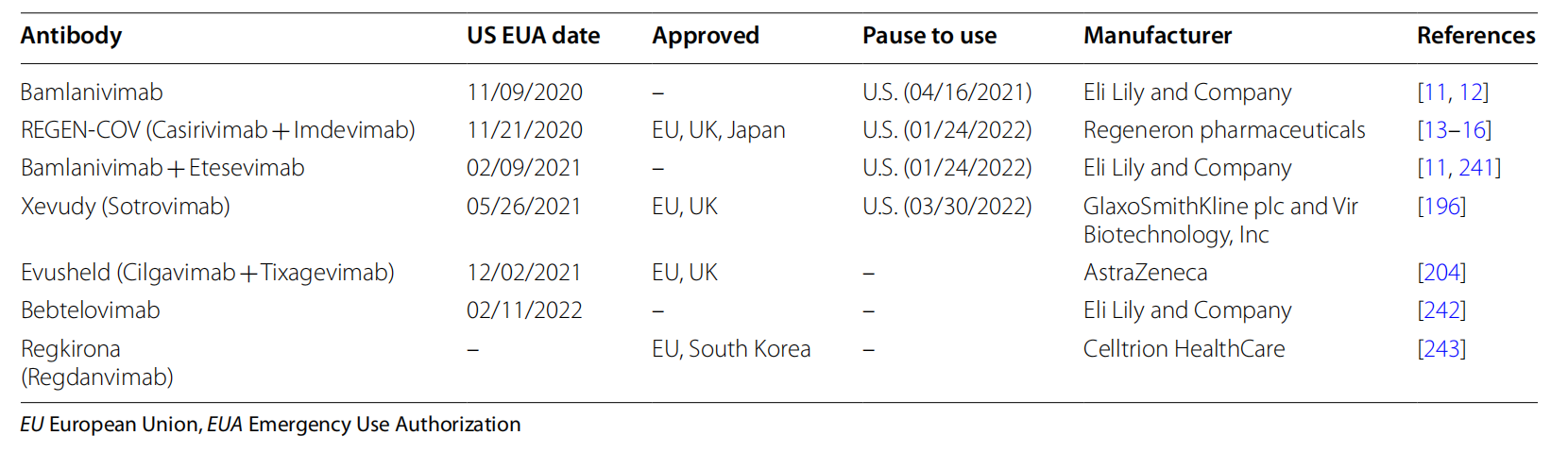
In September 2021, the U.S. FDA approved the Emergency Use Authorization (EUA) for the combination therapy of bamlanivimab and etesevimab developed by Eli Lilly and Company. Bamlaniimab and etesevimab are approved for intravenous infusion in adult and pediatric patients aged 12 years and older, as well as elderly patients with mild to moderate COVID-19 symptoms. Both bamlanivimab and etesevimab are prepared by isolating antigen-specific B cells from recovered COVID-19 patients; the two nAbs target different but overlapping epitopes within the SARS-CoV-2 S protein RBD.
Sotrovimab was developed by GlaxoSmithKline and Vir Biotechnology. This nAb was isolated from memory B cells of patients infected with SARS-CoV-1. This monoclonal antibody does not block the interaction between the viral S protein and the host ACE2 receptor; instead, it targets a common RBD epitope of sarbecovirus, thus neutralizing multiple variants, including Beta, Gamma, Delta, and Omicron BA.1.

(Data source: Kumari M, et al. J Biomed Sci. 2022)
During the COVID-19 pandemic, numerous combination therapies have emerged, some of which aim to block the binding of SARS-CoV-2 to its receptors, thereby inhibiting the virus from entering human cells. These therapies typically combine antiviral drugs with immunomodulators, targeting multiple disease mechanisms to improve treatment outcomes.
IBIO123 is a hybrid monoclonal antibody developed by Immune Biosolutions designed to neutralize the SARS-CoV-2 virus by targeting its S protein and preventing entry into human cells. The therapy consists of three fully human recombinant monoclonal IgGs: IBIO-1 (63%), IBIO-2 (5%), and IBIO-3 (32%). IBIO123 is formulated as an inhaler and delivered directly to the lungs, where it exerts a local effect to neutralize the virus. IBIO-1 and IBIO-3 bind non-competitively to the RBD, while IBIO-2 binds to a highly conserved epitope in the S2 subunit. Results from Phase I and II clinical trials (NCT05639166) have shown reduction in respiratory symptoms without significant safety concerns. Therefore, IBIO123 shows promise in symptom relief, making it a promising candidate treatment for symptomatic COVID-19 patients.
Ronapreve, also known as REGN-COV, was developed by Roche and Regeneron Pharmaceuticals and is based on a combination of two non-competitive monoclonal antibodies (casirivimab and imdevimab). These monoclonal antibodies bind to non-overlapping regions of the SARS-CoV-2 S protein RBD, preventing the virus from entering human cells by inhibiting its binding to the ACE2 receptor. It has shown efficacy in reducing viral load, alleviating symptoms, and significantly reducing the risk of COVID-19-related hospitalization or death. A Phase III clinical trial, NCT04852978, aimed to evaluate the safety and immune response of Ronapreve in combination with Moderna's mRNA-1237 vaccine in individuals with chronic diseases.
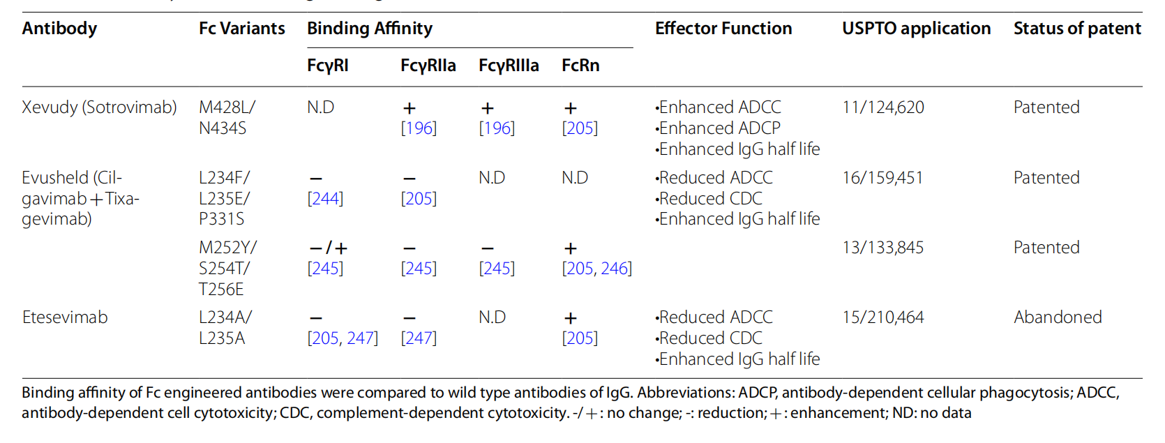
(Data source: Kumari M, et al. J Biomed Sci. 2022)