Escherichia coli (EHEC) and Shigella serotype 1 are enterohemorrhagic bacilli that cause hemorrhagic colitis. This can lead to potentially fatal complications such as hemolytic uremic syndrome (HUS), characterized by thrombocytopenia, acute renal failure, and neurological abnormalities. Both bacteria produce Shiga toxin (Stx), an exotoxin encoded by bacteriophages that inhibits protein synthesis in host cells and is primarily responsible for bacterial virulence.
Shiga toxin structure
The toxins produced by bacteria, specifically enteropathogenic Escherichia coli (EHEC) and Shigella dysenteriae serotype 1, are the main virulence factors. Two major structural types of Stxs have been identified: Shigella toxin type 1 (Stx1) and Shigella toxin type 2 (Stx2). These are further subdivided into subtypes: Stx1a, Stx1c, Stx1d; Stx2a, Stx2b, Stx2c, Stx2d, Stx2e, Stx2f, Stx2g, Stx2h, and Stx2i. Each subtype contains multiple variants released by Stx subtype-specific STEC strains.
Stx is an AB5 toxin composed of an enzymatically active A subunit (Stx-A, 32 kDa) and five identical B subunits (Stx-B, 7.7 kDa), forming a pentameric structure. The A subunit non-covalently binds to the pentamer by inserting its C-terminal region into the central pore. The cytotoxic activity of the A subunit is associated with an RNA N-glycosidase that cleaves 28S ribosomal RNA, thereby inhibiting protein synthesis. The B subunits are responsible for binding to a receptor.

(Data source: Lee KS, et al. Toxins (Base)l. 2021)
The sphingolipid glycolipid ceramide (Gb3, also known as CD77) is the major receptor for Stx. After Gb3 accumulates on the cell surface, Stx is internalized via clathrin-dependent or clathrin-independent endocytosis, then retrogradely sequenced into the trans-Golgi network (TGN), and further into the endoplasmic reticulum (ER), bypassing the late endocytosis pathway. The A subunit is further processed by host furin proteins and furin-like proteases (cleaved between R251 and M252 positions of Stx1 and between R250 and A251 positions of Stx2), forming the enzymatically active A1 fragment (27.5 kDa) and the linker A2 fragment (4.5 kDa), which are linked to their B subunits. The A1 and A2 domains are linked by a single disulfide bond, which is reduced in the ER. The A1 amino acid is then released from the ER into the cytoplasm via the ER-associated protein degradation (ERAD) pathway to inhibit protein synthesis. In particular, the B subunit mediates the specific binding of the toxin to the target cell receptor and promotes the entry of the toxin into the target cell via receptor-mediated entry.
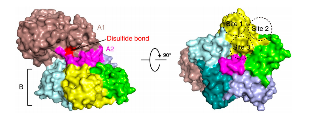
(Data source: Liu Y, et al. Toxins (Basel). 2021)
The role of Shiga toxin in immune regulation and disease
STEC induces the release of Shiga toxins and the production of cytokines and chemokines through colonization in intestinal epithelial cells. In addition to Stxs-induced cell death, various cells, including chemotactic-induced neutrophils, trigger inflammation in the gut, leading to damage. The toxins cross the intestinal mucosa, enter the bloodstream, and reach target organs such as the kidneys and central nervous system. Following membrane-invasive endocytosis mediated by the cell surface toxin receptor Gb3, Stxes migrate to the Golgi apparatus and the ER. Shiga toxins, as a multifunctional bacterial protein, promote ER stress, ribosomal toxicity stress, pro-inflammatory responses, apoptosis, and autophagy in host cells.
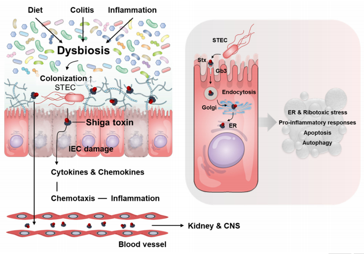
(Data source: Lee KS, et al. Toxins. 2021)
Hemolytic uremic syndrome (HUS) is a group of clinical disorders characterized by low levels of red blood cells and platelets, and acute kidney injury (AKI). The pathological manifestation of HUS is thrombotic microangiopathy. Shiga toxin-producing Escherichia coli (STEC) is the main pathogen causing typical and diarrhea-associated HUS (D+HUS). When food or water contaminated with STEC is ingested, STEC colonizes the colonic epithelium. Shiga toxins (Stxs), the main virulence factor of STEC, are released from STEC, further disrupting the vascular network of the intestinal mucosa, leading to hemorrhagic colitis. Once Stx enters the systemic circulation, it can bind to leukocytes and platelets in the blood and be transported to the kidneys and other target organs. Stxs can bind to glycolipid sphingosyltrihexose (Gb3Cer) on the cell membrane surface and induce endocytosis. Then, Stxs are transported to the opposite side of the Golgi network via the reverse transport pathway, reaching the endoplasmic reticulum (ER). In the ER, the enzyme domains of Stxs are released into the cytoplasm, inhibiting protein synthesis and leading to apoptosis, ER stress, inflammation, and damage.
Stx2 can also bind to monocytes, leading to the release of cytokines such as interleukin-1 (IL-1) and tumor necrosis factor (TNF-α). These cytokines can upregulate Gb3Cer expression in endothelial cells. Bacterial endotoxins such as lipopolysaccharides ( LPSs ) can also enhance Stx2-triggered TNF-α production, which facilitates neutrophil adhesion to the vascular endothelium and the release of inflammatory mediators, further exacerbating endothelial cell damage and leading to the loss of their normal physiological functions, including inhibition of thrombus formation, leukocyte adhesion, and complement activation. Stx2 can also cause complement deposition in erythrocytes (RBCs), leading to the release of hemoglobin and LDH (a marker of intravascular hemolysis) in STEC-HUS patients, as well as the release of complement-coated RBC-derived microvesicles in vitro, thereby triggering complement-mediated hemolysis.

(Data source: Liu Y, et al. Toxins (Basel). 2022)
Targeted therapy with Shiga toxin
Several monoclonal antibodies have been developed to neutralize the toxicity of Stxs, such as monoclonal antibodies against Stx1 and 2 (cαStx1 and cαStx2, Shigamabs®), and the humanized monoclonal antibody against Stx2, Urtoxazumab (TMA-15). These monoclonal antibodies have shown promising results in preclinical studies, and their efficacy will be further validated in clinical trials.

(Data source: Liu Y, et al. Toxins (Basel). 2022)
Urtoxazumab (TMA-15) is a humanized monoclonal antibody against Shiga toxin B subunit (Stx)2 developed by Teijin. It protects mice from death within 24 hours of STEC infection and reduces brain damage and death in a probiotic pig model. TMA-15 was found to be well-tolerated in intravenous Phase I and Phase II clinical trials in healthy adults and pediatric patients with confirmed STEC infection.
One study compared two doses of urtoxazumab (3.0 mg/kg and 1.0 mg/kg) with placebo. Whether 3.0 mg/kg urtoxazumab (71 participants: RR 0.34, 95% CI 0.01-8.14) or 1.0 mg/kg urtoxazumab (74 participants: RR 0.95, 95% CI 0.06-14.59) reduced the incidence of HUS compared to placebo is uncertain (very low certainty of evidence). Low-deterministic evidence suggests that the number of treatment-emergent adverse events may be little or no different between placebo and 3.0 mg/kg urtoxazumab (71 participants: RR 1.00, 95% CI 0.84-1.18) or 1.0 mg/kg urtoxazumab (74 participants: RR 0.95, 95% CI 0.79-1.13). It is currently uncertain whether either dose of urtoxazumab increases the risk of neurological complications or death (very low-deterministic evidence).