Ebola virus (EBOV) is defined as a prototypical member of the genus Ebolavirus, which belongs to the family Filamentous Enveloped Nonsegmented Negative-sense RNAviridae and is responsible for sporadic outbreaks in sub-Saharan Africa. EBOV is a highly contagious virus that spreads through infection in humans or animals and causes a nearly fatal disease, Ebola virus disease (EVD). Due to its complex structure and the lack of appropriate and effective vaccines and treatments, EBOV remains a significant public health challenge.
Ebola virus particles enter the human body through skin lesions (microscopic or gross wounds) or direct contact with mucous membranes. The primary targets of infection are macrophages and dendritic cells. Infected macrophages and dendritic cells migrate to regional lymph nodes, simultaneously producing progeny virions. Systemic distribution of progeny virions and secondary target cell infection occur in almost all organs by suppressing innate, adaptive, and intrinsic immune responses. The EBOV trimeric glycoprotein (GP) is the only surface glycoprotein that penetrates the viral envelope to mediate viral entry into cells. Therefore, it is a major antigen in vaccines and a target for protective vaccines and infection-inducing antibodies.
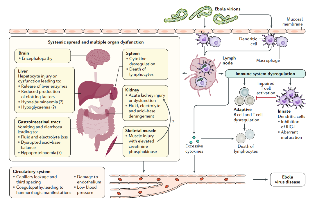
(Data source: Jacob ST, et al. Nat Rev Dis Primers. 2020)
EBOV's structure and function
EBOV is a deadly pathogen that causes severe hemorrhagic fever in humans and non-human primates. Its genome encodes seven structural or multifunctional proteins. The EBOV genome contains approximately 19,000 nucleotides and encodes seven structural proteins. These proteins include an envelope glycoprotein, a nucleoprotein, RNA polymerase L, and viral proteins VP30, VP24, VP35, and VP40.
Ebola glycoproteins are important components of the Ebola virus (EBOV), playing a crucial role in the virus's structure and function. Located on the viral surface, they consist of two subunits , GP1 and GP2. The GP1 subunit is responsible for binding to the host cell and initiating the viral entry process. After binding to the host cell, the GP1 subunit undergoes a conformational change, exposing the GP2 subunit. This allows the virus to fuse with the host cell membrane and release its contents into the cell.
During the viral life cycle, GP undergoes significant conformational changes. Pre-fusion GP is a goblet trimer composed of GP1 and GP2 heterodimers, formed through post-translational cleavage mediated by furin proteases within the host cell; at this stage, the GP polypeptides are still linked by disulfide bonds. GP1 contains domains responsible for cell adhesion and receptor binding; while GP2 contains domains responsible for viral membrane fusion with the endoplasmic reticulum membrane and release of the virus into the cytoplasm. During infection, the mucinous domain (MLD) and glycosyl cap (GC) domain of GP1 are cleaved by trypsin from the host endoplasmic reticulum lysosomes to ensure that the receptor-binding region (RBR) can be recognized by the host receptor NPC1. NPC1, acting as an acidic pH trigger, can, upon binding to other factors (not yet fully understood), induce a significant conformational rearrangement of GP, thereby achieving the fusion process.

(Data source: Misasi J, et al. Immunity. 2021)
Interferon response mechanism and signaling pathway after EBOV infection
Based on the IFNα/β signaling pathway: Activation of tyrosine kinase-related receptors JAK1 and TYK2 via the interferon α receptor (IFNαR) promotes phosphorylation and dimerization of STAT1 and STAT2. IFNγ signaling pathway: IFNγ binding to the IFNγ receptor (IFNγR) activates JAK1 and JAK2, similarly promoting phosphorylation and dimerization of the homodimer STAT1.
EBOV inhibits the expression of IFN-induced genes by competitively binding to the cytoplasmic protein KPNAα (KPNA). Furthermore, EBOV's VP35 inhibits the production of cytokines such as IL-6, IL-12, TNFα, and IFNα/β. When Toll-like receptor 4 (TLR4) detects viral lipopolysaccharide, its activation initiates a downstream signaling cascade, leading to the activation of NF-κB, IRF3, and IRF7. Due to the loss of function of major histocompatibility complex class II (CMH II) in cytokine production and antigen presentation, VP35 further inhibits the phosphorylation of NF-κB, IRF3, and IRF7.

(Data source: Martin Ndayambaje, et al. Egyptian Journal of Medical Human 2024)
Targeted therapy for EBOV
ZMapp, an optimized antibody composed of three monoclonal antibodies (two from ZMAb (2G4, 4G7) and one from MB-003 (13C6)) reversed late-stage EVD in rhesus monkeys and rescued 100% of these animals, even though treatment was given 5 days after the EBOV challenge. In clinical trials, ZMapp's performance varied. One clinical trial involving 169 patients showed an overall mortality rate of 49.7%, with the first negative viral test result appearing in 27 days. This suggests that while ZMapp is highly effective in animal models, its efficacy in humans may be influenced by various factors, such as patient disease severity and timing of treatment.
Two recently developed monoclonal antibody treatments, mAb114 and REGN-EB3, are superior to ZMapp in reducing mortality from EVD and have now been approved by the U.S. Food and Drug Administration as treatments for EBOV infection.

(Data source: Jacob ST, et al. Nat Rev Dis Primers. 2020)
Ebanga® (ansuvimab-zykl, formerly mAb114) is a recombinant human monoclonal antibody for the treatment of infections caused by the Zaire Ebola virus in adults and pediatric patients, including newborns whose mothers are infected with the Zaire Ebola virus and who test positive for RT-PCR. On December 21, 2020, Ridgeback Corporation received FDA approval for Ebanga®.
Inmazeb, formerly known as REGN-EB3, was developed using Regeneron's VelocImmune® platform and related VelociSuite® technology and has received FDA approval. This therapy consists of three structurally similar monoclonal antibodies: atolativimab, maftivimab, and odesivimab, which bind to different, non-overlapping epitopes on Ebola virus glycoproteins. REGN3479 (maftivimab) targets the fusion loop and has potent neutralizing activity. REGN3471 (odesivimab) targets the GP1 head and sGP. Although REGN3471 has weaker neutralizing ability, it mediates effector function and provides partial protection in an EBOV-infected guinea pig model. REGN3470 (atolativimab) targets the glycosyl cap, has partial neutralizing activity, and mediates Fc effector function, which can promote the killing of EBOV-infected cells. These three antibodies help neutralize the Ebola virus by blocking its entry into the patient's body (and/or) or by mobilizing other immune cells to target and eliminate infected cells. Inmazeb can prevent the rapid emergence of escape mutants, something that monotherapy, even targeting conserved epitopes, cannot do.


(Data source: Rayaprolu V, et al. Cell Host Microbe. 2023)