Rabies virus (RABV) is a single-stranded, negative-sense RNA virus belonging to the family Rhabdoviridae and the genus Rabievirus. RABV is a highly neurotropic virus that can infect almost all warm-blooded animals, including humans. RABV primarily infects neurons, but there is evidence that it can also infect astrocytes and microglia. Rabies virus can cause fatal viral encephalitis in various hosts. Prevention relies on timely post-exposure prophylaxis (PEP) after the onset of symptoms, including vaccination and administration of rabies immunoglobulin (RIG) to obtain immediate immune protection. While vaccines and immunoglobulins can prevent the development of post-exposure rabies, there is currently no effective drug treatment for rabies. The high mortality rate of rabies poses a serious threat to human health and public safety.
Structural components and functions of RABV
Rabies virus (RABV) belongs to the family Rhabdoviridae and the genus Rabievirus. It is a single-stranded negative-sense RNA virus with a genome length of approximately 12 kb, encoding five proteins: nucleoprotein (N), phosphoprotein (P), matrix protein (M), glycoprotein (G), and RNA polymerase (L).
The N protein of rabies virus encapsulates the RNA genome, forming a tightly bound N-RNA complex called a ribonucleoprotein body (RNP). The RNP, along with the L and P proteins, forms a helical nucleocapsid (NC), which serves as a template for rabies virus RNA transcription and replication. The P protein of rabies virus is a catalytic cofactor of polymerase L and can disrupt the host's interferon-mediated antiviral response.
The RABV M protein encapsulates the NC and is located between the N and G proteins. It interacts with the transmembrane domains of the G protein and RNP, participates in viral budding and RNA replication, and connects the NC to the viral envelope.
The RABV G protein is a trimer and the only protein exposed on the naked viral membrane surface. Its extracellular region is involved in cell receptor recognition, adhesion, and viral invasion. The G protein has also been shown to be closely associated with neuronal apoptosis. In the early stages of rabies virus (RABV) infection, the virus is retrogradely transported along axons through the neuromuscular junction to reach the central nervous system. The G protein is a major target of virus-neutralizing antibodies (VNAs), providing protection against rabies. It interacts with host cell receptors and promotes pH-triggered fusion, releasing RNPs into the host cell cytoplasm.

(Data source: Kiflu AB. Viruses. 2024)
RABV inhibits interferon response
RABV infection activates the interferon signaling pathway, while RABV inhibits this pathway's activity by interacting with key factors within the pathway through its own proteins. The RABV P protein directly binds to tyrosine-phosphorylated STAT (pY-STAT), affecting its localization and reducing its ability to bind to the ISG (interferon-stimulated gene) promoter, thereby inhibiting ISG expression. Furthermore, the P protein interacts directly with TBK-1 (TANK-binding kinase-1) in a dose-dependent manner, inhibiting IRF3 (interferon regulatory factor 3) phosphorylation. Simultaneously, the interaction between the P protein and PML (promyeloid leukemia-associated protein) alters the localization of PML protein and the structure of the PML nucleosome, thereby regulating IFN-induced apoptosis. The encapsulation of RABV RNA by the N protein prevents viral RNA from being recognized by RIG-I (retinoic acid-inducible gene I), thus preventing RIG-I-mediated activation of the downstream IRF-3 pathway. Type I IFN stimulation causes the M protein to transition to an activated pSTAT1-interacting state, thereby enhancing the ability of the P protein to bind to JAK1, preventing pSTAT1 from being activated, and inhibiting it in the cytoplasm through interaction with pSTAT1.

(Data source: Zhang H, et al.Animal Model Exp Med. 2022)
The role of RABV in autophagy and apoptosis
RABV can activate the initiation of autophagy. After viral invasion, RABV activates the AMPK signaling pathway. On the one hand, activated AMPK inhibits mTORC1, thereby relieving the mTORC1 inhibition of the ULK1 complex; on the other hand, AMPK can positively regulate the AKT and MAPK factors, promoting the activation of the ULK1 complex and thus driving the initiation of autophagy. Furthermore, the P protein of rabies virus binds to Beclin1 in the PI3KC3 complex, reducing the phosphorylation level of CASP2. This not only positively regulates the AMPK signaling pathway but also negatively regulates the mTORC1 signaling pathway, thereby activating the initiation of autophagy. RABV prevents the fusion of autophagosomes and lysosomes. The P protein of RABV binds to Beclin1, encapsulating immature autophagosomes, inhibiting the fusion of autophagosomes and lysosomes, and blocking the degradation of autophagosomes. IFIM3 can directly inhibit the ULK1 complex, promote the phosphorylation of mTORC1, and indirectly inhibit the ULK1 complex, thereby blocking the initiation of autophagy caused by RABV infection.

RABV can induce mitochondrial apoptosis in the late replication phase, and its regulation is controlled by the BCL-2 gene family. Three RABV proteins, M, G, and P, are closely related to apoptosis. The M protein can inhibit the anti-apoptotic gene Bcl2, thereby leading to mitochondrial apoptosis and the release of cytochrome C and AIF factors.
Caspase-dependent pathway: Released cytochrome C activates caspase-9, which in turn activates downstream caspase-3, inducing apoptosis. Non-caspase-dependent pathway: Released AIF can directly induce apoptosis.

(Data source: Li S, et al. Cells. 2024)
Targeted therapy for RABV
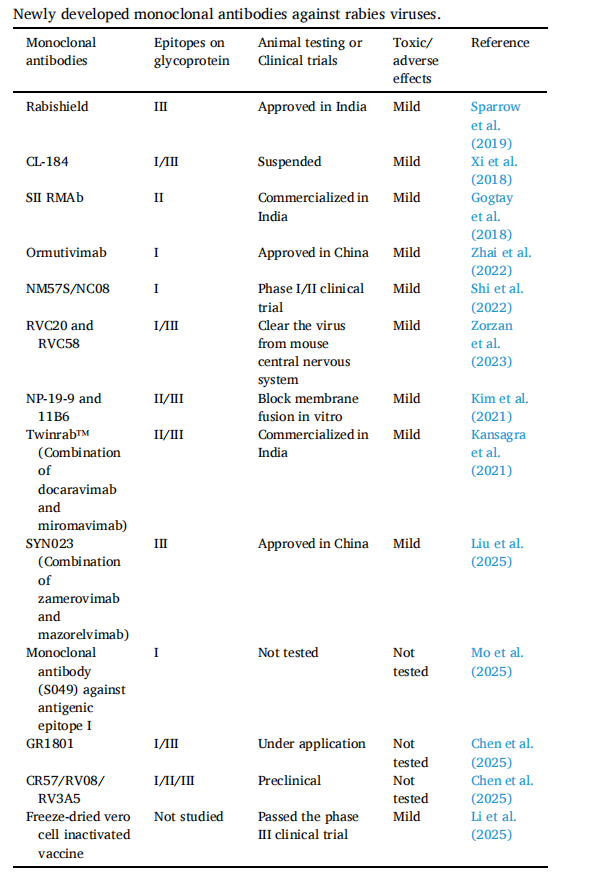
Rabies glycoprotein (RVGP) is commonly used as a target antigen in rabies vaccine development. This protein is the only viral protein found on the surface of RABV particles. RVGP is essential for RABV infection of host cells and is associated with the virus's cytotropism and virulence. The protective efficacy of vaccines and immunoglobulins is directly related to the production of neutralizing antibodies against RVGP. RVGP contains four major antigenic sites : site I (residues 226-231), site II (residues 34-42 and 198-200), site III (residues 330-338), and site IV (residues 261-264). Among these, sites II and III are the primary targets for neutralization, and approximately 90% of known monoclonal antibodies target them.
(Data source: Yuan S, et al.) Antiviral Res. 2026)
SYN023 consists of two human IgG monoclonal antibodies, CTB011 (zamerovimab) and CTB012 (mazorelvimab). These antibodies bind to disjoint, non-overlapping epitopes located in highly conserved regions of the rabies virus glycoprotein (RVGP). CTB011 targets a peptide near antigenic site iii, while CTB012 targets a discontinuous but highly conserved region within a known antigenic site that is not classified. Mouse model studies have shown that the two antibodies provide cross-protective neutralization against escape mutants generated during infection when administered continuously. Clinical studies in patients exposed to rabies virus have further evaluated SYN023 as part of post-exposure prophylaxis, comparing its efficacy and safety with human rabies immunoglobulin. Results suggest that this monoclonal antibody combination provides effective protection more quickly after exposure compared to conventional RIG. SYN023 has successfully completed Phase 3 clinical trials and is expected to become the first commercially licensed humanized rabies virus antibody cocktail. The combination therapy of zamerovimab and mazorelvimab was approved by regulatory authorities in China in 2024. The efficacy of antibodies against rabies PEP may be affected by RVGP mutations. Sites II and III are the main targets for neutralization. However, unlike the highly conserved and immutable antigenic site I, these epitopes are prone to mutation. Leveraging this stability, recombinant RVGP was redesigned and expressed in 293FT cells to isolate a site I-specific monoclonal antibody (S049) (therapeutic data are currently unavailable). This strategy of targeting non-mutated epitopes provides a promising approach to avoiding immune escape.

(Data source: Cao L) et al. Emerg Microbes Infect. 2025)
Silevimig is a bispecific antibody targeting the RABV glycoprotein (RVGP), simultaneously and specifically binding to two non-overlapping epitopes (epitaphth I and epitope III) on the G protein. Epitope I arm is a single-chain variable domain, and epitope III arm is a Fab domain. A heterodimeric monoclonal antibody is formed by introducing a button-in-place pore (KIH) technique into the Fc domain. In vitro studies have shown that Silevimig inhibits 90 rabies virus G protein mutations, 21 pseudotypes of rabies virus, and 18 wild-type rabies virus.

(Data source: Long C, et al. J Med Virol. 2023)