Methamphetamine(METH), also known as methylamphetamine or desoxyephedrine, is commonly called "ice" because it appears as colorless, transparent crystals. Methamphetamine is a novel psychoactive substance with complex neurotoxicity, severely affecting the central nervous system (CNS). Methamphetamine (METH) use induces systemic inflammation, characterized by elevated levels of inflammatory factors. Methamphetamine use disorder (MUD) is characterized by compulsive drug use and significant neurotoxicity, imposing a heavy burden on individuals and society. Traditionally considered a localized CNS disorder, recent preclinical and clinical studies have elucidated that MUD is a multidimensional disorder affecting multiple biological systems, particularly the immune system.
The structure and receptor of METH
Methamphetamine (N-methyl-α-methylphenethylamine) is a cationic molecule and a chiral compound based on the phenethylamine core structure. Its significant difference from its amphetamine analogues lies in the presence of a methyl group. This functional group gives methamphetamine strong lipophilicity, allowing it to penetrate the blood-brain barrier more effectively. In neural tissue, methamphetamine promotes catecholamine signaling (such as dopamine and norepinephrine) through multiple mechanisms. It binds to vesicle monoamine transporter-2 (VMAT2) and accumulates within vesicles, altering the pH and leading to the release of catecholamines into the cytoplasm.
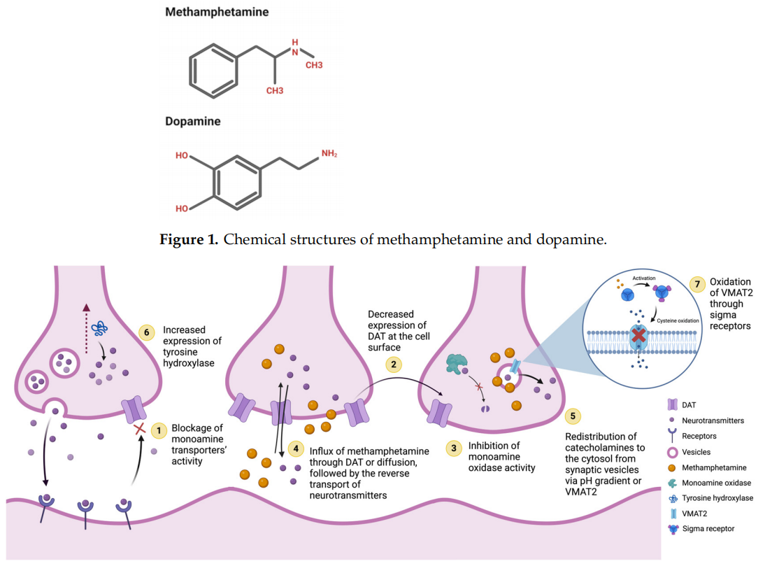
(Data source: Khan R, et al. Biomolecules. 2025)
The role of METH in neuroinflammation and immune regulation
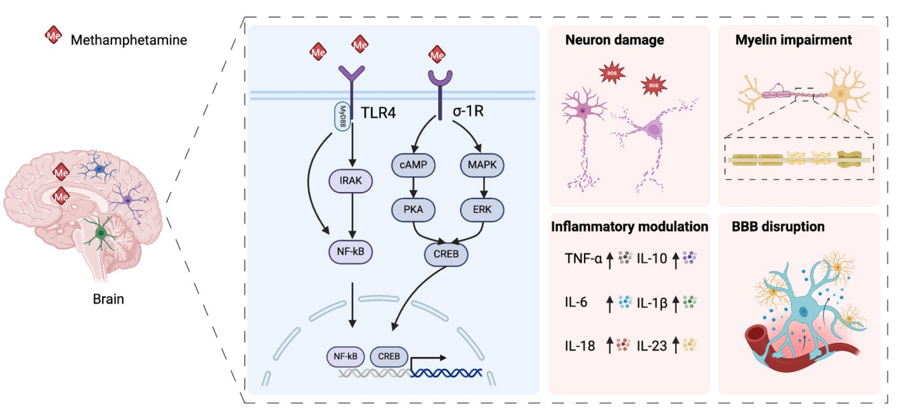
The effects of methamphetamine (METH) on the central nervous system (CNS) are mediated through complex cellular and molecular pathways, including the activation of resident immune cells and the subsequent release of inflammatory mediators. Growing evidence suggests that glial cells, including microglia, astrocytes, and oligodendrocytes, are activated in the CNS during methamphetamine use disorder (MUD).
METH exposure activates astrocytes via TLR4 and σ-1R, which in turn triggers downstream responses through the MyD88/IRAK/NF-κB, cAMP/PKA, and MAPK/ERK signaling pathways. This activation leads to the release of inflammatory cytokines, disruption of the blood-brain barrier, myelin dysregulation, and neuronal damage.
METH exhibits toxic effects on immune organs, including the liver, spleen, intestines, and lymph nodes. METH exposure is associated with increased steatosis, fibrosis, and necrosis, and can lead to liver lobule degeneration. METH upregulates the activity of Kupffer cells and stellate cells through the TLR4 and PI3K/AKT signaling pathways, thereby increasing levels of FGF, GPx-1, SOD-1, and BA. Furthermore, METH affects antigen presentation by reducing the ability of macrophages to activate CD4+ and CD8+ T cells, and promotes the production of pro-inflammatory cytokines such as IL-1β and TNF-α by NK cells. METH exposure leads to splenomegaly and impaired filtration function. Additionally, it can induce T cell and splenocyte apoptosis and inhibit IL-2 production. METH exposure also promotes the production of IFN-γ, TNF-α, IL-6, and IL-12 by activating T cells, NK cells, and macrophages. METH exposure induces acute ischemia and disrupts barrier permeability. Simultaneously, it upregulates LPS levels and downregulates the production of short-chain fatty acids (SCFAs) via the TLR4 signaling pathway, leading to endotoxemia. Furthermore, methamphetamine (METH) exposure causes immunosuppression, manifested as downregulation of dendritic cells (DCs) and natural killer cells (NK cells). It also upregulates pro-inflammatory cytokines, including IL-1, IL-6, IL-8, and TNF-α released by macrophages and CD4+ T cells. METH also induces lymph node enlargement via the TAAR1/CREB signaling pathway and promotes the upregulation of IL-2.

Methamphetamine addiction causes systemic damage through three potential mechanisms. First, methamphetamine exposure can activate the reticular formation pathway (NTS) and dorsal vagal nucleus (DMV) of the vagus nerve, leading to the release of acetylcholine and norepinephrine. This activation can impair the function of peripheral immune organs, including the intestines, liver, and spleen, potentially creating a positive feedback loop. Second, long-term methamphetamine exposure can activate the hypothalamic-pituitary-adrenal (HPA) axis, inducing the release of cortisol, thereby causing immunosuppression and constituting a negative feedback mechanism. Third, methamphetamine can directly or indirectly damage the blood-brain barrier (BBB), activate immune cells, and promote the production of cytokines and chemokines, which can directly affect peripheral organs.
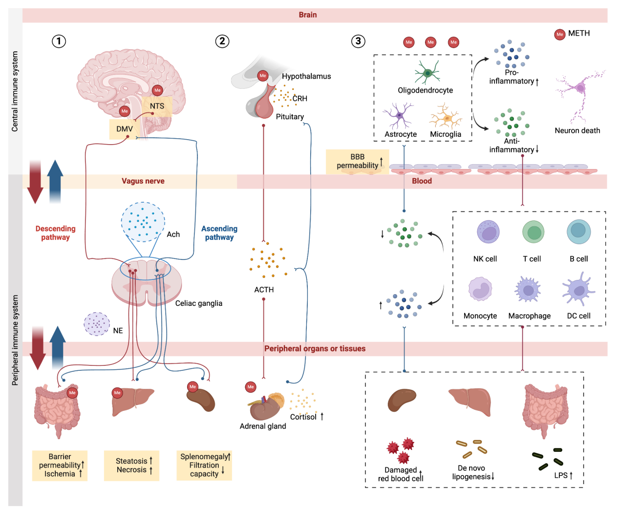
(Data source: Shi S, et al. J Neuroinflammation. 2025)
METH targeted therapy
Devextinetug (IXT-M200) is a monoclonal antibody targeting methamphetamine. In a phase II human trial called "STAMPOUT" (Ephedrine Outpatient Treatment Antibody Study, NCT03336866), in a non-seeker population, IXT-m200 administration significantly reduced the volume of distribution of methether in the blood within up to 3 weeks after an injection of 30 mg of methether. No serious adverse events following IXT-m200 administration were observed in the STAMPOUT program. IXT-m200 shows potential in the treatment of methfelter-dependent dysplasia (MUD).
Devextinetug possesses a mouse-derived variable binding region, termed "7F9", and a humanized constant domain. Humanization of the variable region of ch-mAb7F9 through multiple sequence manipulations yielded 48 IgGs. Eight candidate IgGs (including the parental IS12) were selected for further testing. Ligand cross-reactivity and performance in a METH-induced rodent motor stimulation model were tested on these eight IgGs. Cross-reactivity results showed that IS12 exhibited the strongest affinity for methamphetamine (METH) and, compared to the other seven candidates, demonstrated the greatest reduction in METH-induced activity enhancement.

(Data source: Berquist MD, et al. Psychopharmacology (Berl). 2015)