Phosphatidylserine (PS) is an anionic phospholipid found on the membranes of various organelles within cells, most notably the plasma membrane. Under homeostatic conditions, PS is normally confined to the inner layer of the plasma membrane. However, during cell activation and/or cell-induced death, PS is everted to the outer surface of the plasma membrane via activation by phospholipid eversion enzymes. This everted PS not only alters the biochemical and physical properties of the plasma membrane but also initiates a series of interactions between endogenous extracellular proteins and receptors on neighboring cells to stimulate phagocytosis (paracellular phagocytosis), thereby influencing the surrounding immune environment. PS is an active immunomodulatory molecule in the tumor microenvironment; in this environment, aberrantly exposed PS can mediate immunosuppression, helping tumor cells evade the surveillance of the immune system.

(Data source: Calianese DC, et al. Cell Commun Signal. 2020)
The molecular structure and receptor of Phosphatidylserine
Phosphatidylserine (PS) consists of a glycerol backbone esterified at the sn-1 and sn-2 carbons of the glycerol moiety, two fatty acyl chains of variable length and saturation, and a phosphate bond covalently linked to a serine residue at the sn-3 position. In eukaryotic cells, PS is synthesized from phosphatidylcholine (PC) and phosphatidylethanolamine (PE) by PSS1 and PSS2, respectively, and can be catabolized by phosphatidylserine decarboxylase (PSD) to produce PE.

(Data source: Birge RB , et al. Cell Death Differ. 2016)

(Data source: Chang W, et al. Theranostics. 2020)
Immune cells possess a variety of PS receptors. The mechanisms by which these receptors recognize PS are crucial for the endocytosis process of immune cells. The PS receptor family is diverse, with two identified as PS- sensing receptors: the TAM receptor protein tyrosine kinase family (TYRO3, AXL, and MER) and the TIM family (T cell/transmembrane, immunoglobulin, and mucin). Other receptors on immune cells include αvβ3 or αvβ5 integrins, CD300a, BAI1, Stabilin, RAGE , and LOX-1.

(Data source: Wang J, et al. Biomark Res. 2022)
TAM binds to PS via proteins containing the Gla domain (such as Gas6 or Pros1). Ca2+ is also involved in efficient PS binding and receptor activation. PS binds to TAM to regulate feedback inhibition of the innate immune response in immune cells. The TAM protein forms a narrow cavity or depression to which PS binds and plays a key role in regulating the immune response. Stabilin-1/2 binds to PS through four clusters (each cluster including an EGF-like domain, an atypical EGF-like domain, a FAS domain, and/or a linker domain), thereby activating a series of signals that lead to immunosuppression.
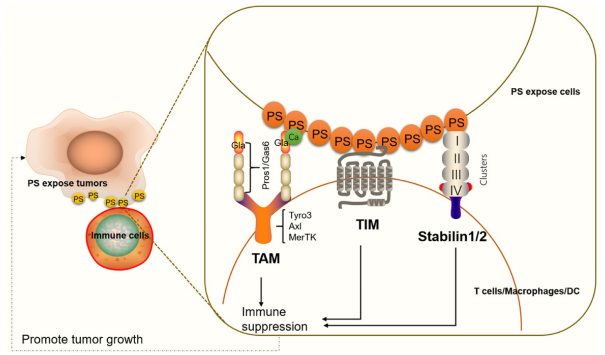
(Data source: Chang W, et al. Theranostics. 2020)
Biological mechanisms of Phosphatidylserine exposure
In normal cells, the PS ( phospholipid reversal enzyme) is maintained in the inner layer of the plasma membrane by translocases such as P4-ATPases and phosphatidylamide reversals (ATP11A and ATP11C). Under apoptosis or strong activation signals, caspase-mediated translocase cleavage or Ca2+ influx disrupts membrane asymmetry and activates exchange enzymes, including Xk-associated protein 8 (Xkr8) and transmembrane protein 16F (TMEM16F), leading to rapid PS eversion.

(Data source: Wang J, et al. Biomark Res. 2022)
The role of phosphatidylserine in tumor immunity
Phosphatidylserine (PS) exposure on tumor cells and vesicles can induce immunosuppression. PS on the surface of tumor cells triggers immunosuppression by binding to receptors on macrophages and T cells. PS binding to PSRs on macrophages promotes the maturation of M2-like macrophages, which secrete the anti-inflammatory cytokines IL-10 and TGF-β. IL-10 and TGF-β are immunosuppressive cytokines that inhibit T cell activation. PS binding to T cell surface receptors can also inhibit T cell activation through G protein-coupled receptor 174 (GPR174)-mediated M2-type macrophage maturation. Conversely, PS exposed on the surface of tumor-derived microvesicles (i.e., PS exfoliated on microvesicles) can promote the clearance of apoptotic cells through phagocytosis, thereby preventing unwanted inflammatory responses and maintaining an anti-inflammatory state in the tumor microenvironment.
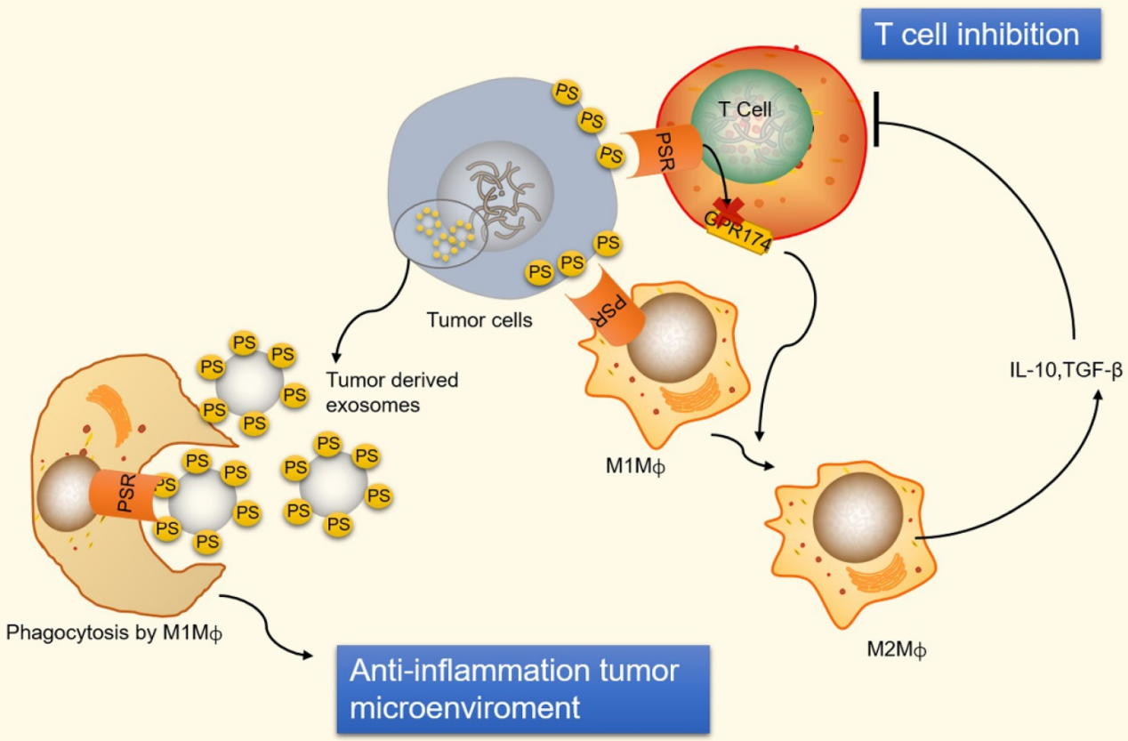
(Data source: Chang W, et al. Theranostics. 2020)

(Data source: Birge RB , et al. Cell Death Differ. 2016)
Targeted therapy with Phosphatidylserine
Chronic exposure to exogenous PS (protein glycosylation) in tumor cells, stromal cells, and tumor-associated blood vessels offers unique opportunities for targeted therapy. Unlike classic immune checkpoints that primarily regulate T cell activation, PS-induced immunosuppression integrates innate, adaptive, and stromal signaling. Therapeutic platforms targeting PS include monoclonal antibodies, nanovesicles, engineered PS-binding proteins, immunomodulatory adjuvants, and emerging cell therapies.
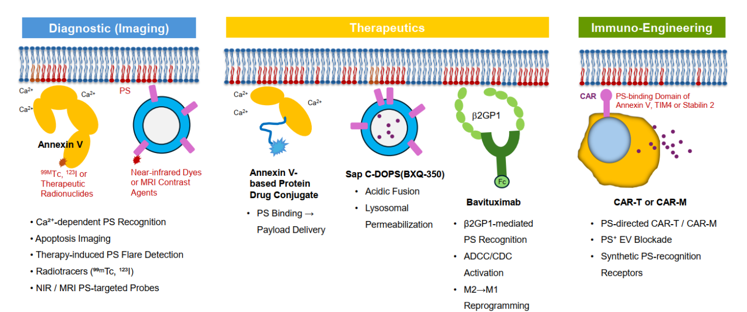
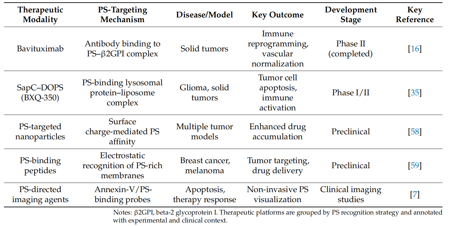
(Data source: Yoo M, et al. Int J Mol Sci. 2026)
Bavituximab is a monoclonal antibody targeting phosphatidylserine, developed by Peregrine Pharmaceuticals for the treatment of non-small cell lung cancer (NSCLC). Bavituximab binds to β2-glycoprotein I (β2GP1), which binds to PS exposed on the surface of tumor cells and compressed endothelial cells. This PS - β2GP1 complex induces FcγR crosslinking and antibody-dependent cytotoxicity, contributing to the remodeling of the immunosuppressive tumor microenvironment. Upon binding, bavituximab can repolarize tumor-associated macrophages to a pro-inflammatory phenotype and promote dendritic cell maturation and antigen presentation. These effects support enhanced natural killer and T cell-mediated antitumor responses. Phase I studies demonstrated a favorable safety profile for bavituximab, with manageable infusion responses and manageable immune-related adverse events. In earlier studies of NSCLC and hepatocellular carcinoma, bavituximab in combination with chemotherapy showed encouraging response signals compared to historical control groups. NCT03519997 was a phase 2 clinical trial investigating the combination of bavituximab (an antibody targeting phosphatidylserine) and pembrolizumab (an anti-PD-1 inhibitor) for the treatment of unresectable hepatocellular carcinoma. The study achieved its predetermined endpoint, demonstrating a confirmed objective response rate (ORR) of 32.1% (9 out of 28 evaluable patients). Treatment-related adverse events occurred in 45.7% of patients, with 14.3% experiencing grade 3 or higher adverse events. B-cell depletion, intratumoral fibrosis, matrix immune checkpoint expression, and immune-rich inflammatory tumors were associated with tumor response.

(Data source: Hsiehchen D, et al. Nat Commun. 2024)